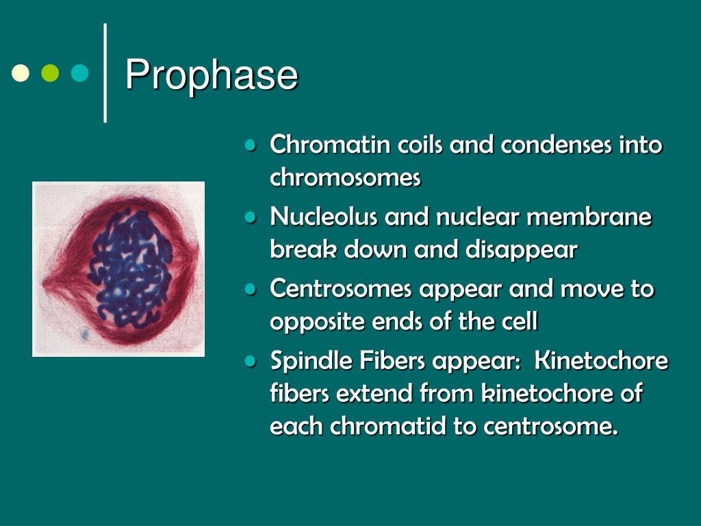
Chapter 121 122 The Cell Cycle Cell Growth Cell Division Biology Diagrams Prophase. During prophase the nucleoli disappear and the chromatin fibers thicken and shorten to form discrete chromosomes visible with the light microscope. Each replicated chromosome appears as two identical chromatids joined at the centromere.. The chromatids shorten and thicken and become more tightly coiled; the individuality of the separate chromosomes becomes clear. During prophase, the chromatin condenses into visible chromosomes. Each chromosome consists of two sister chromatids. These are connected at a region called the centromere. The mitotic spindle also begins to form during this stage. Understanding the carbon cycle is essential for addressing climate change and sustaining ecosystems. Through

Explore the essential stages of prophase in mitosis, focusing on chromosome condensation, spindle formation, and nuclear envelope breakdown. Specific chemical changes to histones, such as phosphorylation, play a role in altering the chromatin structure, making it more compact. These modifications are tightly regulated by various enzymes
Prophase - an overview Biology Diagrams
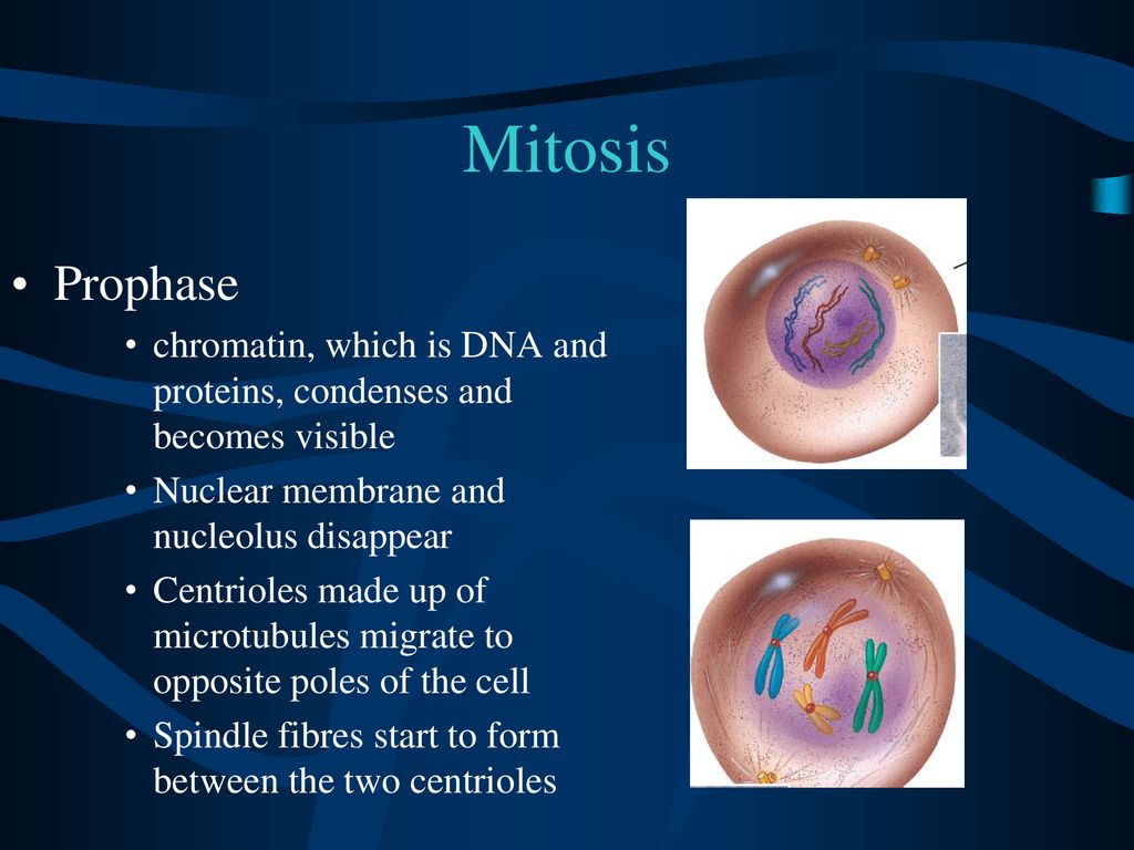
In prophase, the chromatin condenses into discrete chromosomes. The nuclear envelope breaks down and spindles form at opposite poles of the cell. Prophase (versus interphase) is the first true step of the mitotic process. During prophase, several important changes occur: Chromatin fibers become coiled into chromosomes, with each chromosome Early prophase chromatin undergoes an orderly transition as proteins leave chromatin in successive waves that form relatively tight clusters in our analysis. We expected that these early events might involve chromatin changes required to shape mitotic chromosomes. Indeed, HMGN1 and HMGA1 are two of the earliest proteins to leave chromatin. The process is longer due to the phases of prophase which takes place in two phases i.e prophase I and prophase II. Prophase I is quite complex which involves the pairing up of the homologous chromosomes and the exchange of genetic information. It defines the difference between mitosis and meiosis. Prophase II is very similar to the mitotic

phase chromatin changes occur at nuclear pores, on the inner surfaceofthenuclearenvelope, andmoststrikinglyinthenucle-olus. There, proteins involved in rRNA processing move away their proximity to chromatin during prophase. However, about 20% remain relatively unaffected (Figures 3D and 3E; orange cluster). This behavior was well reproduced
Prophase in mitosis and meiosis (Prophase 1 and 2) Biology Diagrams
During prophase, the replicated chromosomes (each consisting of two sister chromatids) condense and are recognizable under the microscope. Survivin is responsible for targeting the CPC to chromatin by associating with histone modifications, Topoisomerase IIα induces a conformational change that places the G-segment within its active
