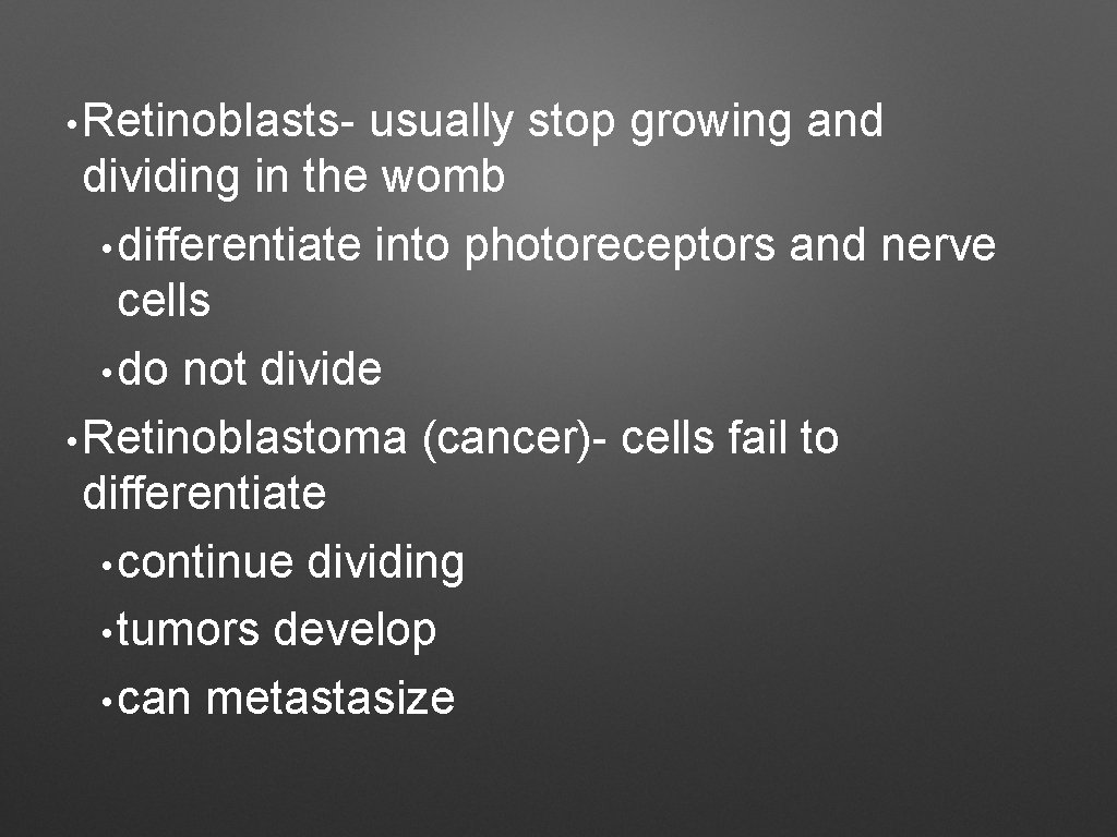
Structure and Function of Tumor Suppressor Uncovered Biology Diagrams Mammalian sterile 20-like kinase 1/2 (MST1/2) are core tumor suppressors in the Hippo signaling pathway. MST1/2 have been shown to regulate mitotic progression. Here, we report a novel mechanism for phospho-regulation of MST2 in mitosis and its biological significance in cancer. We found that the mi … Somatic cells are the best know for this cell cycle and go through 2 main phases called interphase and mitosis. Most cells in the body go through a cycle of life in which their genetic information is retained, fixed, and passed down to daughter cells through a highly coordinated and regulated process. The main tumor suppressor gene that

Mitosis is controlled by a network of kinases and phosphatases. We screened a library of small interfering RNAs against a genome-wide set of phosphatases to comprehensively evaluate the role of human phosphatases in mitosis. We found four candidate spindle checkpoint phosphatases, including the tumor suppressor CDKN3.
Cell Cycle Control, Oncogenes, Tumor Suppressors Biology Diagrams
The stages of the cell cycle (G1: Gap 1, S: DNA synthesis, G2: Gap 2, and M: mitosis) are indicated. Tumor suppressors act to maintain checkpoints (arrows) whereas oncogenes allow for checkpoints Chief among these factors is the drug retention issue, where paclitaxel has been shown to linger in the tumor cells for a week and is thus able to exert its cytotoxicity longer compared with the newer mitosis-selective inhibitors with a median half-life of approximately 13 h. 35, 64, 65, 66 Additionally, it is likely that quiescent cancer cells
Mitosis is controlled by a network of kinases and phosphatases. We screened a library of small interfering RNAs against a genome-wide set of phosphatases to comprehensively evaluate the role of human phosphatases in mitosis. We found four candidate spindle checkpoint phosphatases, including the tumor suppressor CDKN3.

Functional Mechanisms for Human Tumor Suppressors Biology Diagrams
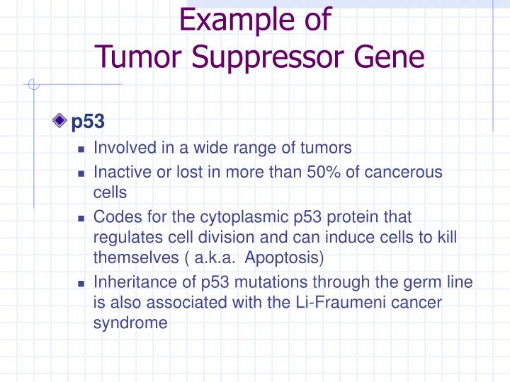
The RB tumor suppressor is well known for its ability to repress transcription and to prevent cell proliferation by arresting cells either in G1, at the G1/S transition, or in S phase of the cell cycle. Other E2F-targets also have well defined roles in mitosis (Table 1, Figure 2) and promote chromosome segregation errors when overexpressed The interference with cell adhesion results in indirect suppression of cell division due to contact inhibition. It has been observed that a few tumor suppressors may act in cooperation to inhibit cell mitosis 3. Tumor suppressors p15, p16, p18, p19, p21 and p27 inhibit cyclin-dependent kinases (CDKs), which, in turn, inhibit Rb 11, 24.